


Insurance Information
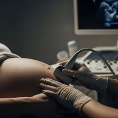
Most health insurance plans will cover some or all OB ultrasounds when medically necessary. Our team is here to help you confirm your coverage and identify any out-of-pocket costs upfront, so there are no surprises.
Patient Resources
From accessing the patient portal to downloading forms, reviewing insurance details, and exploring helpful FAQs and educational guides – we make it easy to stay informed, prepared, and confident in your care.
Meet Our Gynecologists


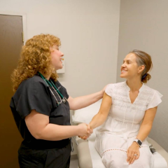
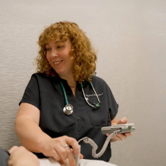
Claire Bareiss, PA-C


.jpg)
.jpg)


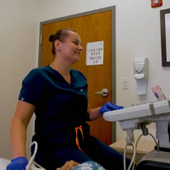
Erin Kopeny, PA-C


Jenny Mathew, WNHP-BC

Madison Monk, PA-C, MS